New imaging technique could provide clearer images for oncologists

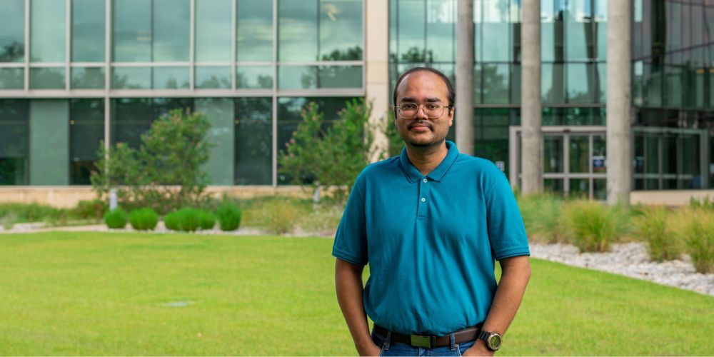
A multidisciplinary team lead by University of Texas at Arlington mathematics Assistant Professor Souvik Roy is on a mission to improve medical imaging using a new technique called quantitative photoacoustic tomography (QPAT).
QPAT is an imaging modality that combines ultrasound, which is an imaging technique that uses sound waves to image features inside the body, and optical tomography. It uses acoustic wave intensity data measured on the surface of the body through a scanner composed of acoustic wave detectors. This allows for the development of images of various optical properties of tissues, like absorption and diffusion, which can provide crucial information regarding the location and stage of a cancerous tissue.
“QPAT is robust because it uses information from two types of imaging techniques and has the potential to provide high-quality images. It can tell us so much more about what’s going on under the skin,” Roy said. “By providing better images, doctors will be able to make more accurate diagnoses in shorter time frames. This will lessen anxiety for patients as well as decrease costs for the health care industry by reducing the need for repeated scans.”
One of the major challenges in developing QPAT is the lack of adequate acoustic wave measurements on the surface of the body, which can significantly degrade the quality of the images and result in inaccurate diagnosis of cancerous tumors. To circumvent this issue, principal investigator Roy and his multidisciplinary team—comprising statistician co-principal investigator Suvra Pal and radiologists—have received a $190,000 grant from the National Science Foundation to significantly develop and improve the QPAT imaging technique.
The team will use a novel combination of game theory, statistical sensitivity analysis and gradient-free optimal control methods to help complete the missing acoustic wave measurements and stabilize and recalibrate computational algorithms for providing high-contrast and high-resolution images.
“We hope to facilitate a safe start to research on imaging human subjects using QPAT,” Roy said. “Our ultimate goal is helping patients get better and develop more accurate images in a shorter time frame. These enhanced scans should help doctors and patients make better health care treatment decisions. Down the line, we know this will improve outcomes, reduce patient anxiety and be highly cost-effective.”