The future of bone healing
A University of Texas at Arlington team researching a groundbreaking treatment to accelerate the healing of cranial injuries has received significant funding support from the National Institutes of Health.
The team, led by Venu Varanasi, is developing a method for live 3D printing of bone-forming scaffolds that can be grafted to cranial defects using regenerative antioxidant materials that support the body’s natural healing processes. The approach could shorten the healing time for severe cranial injuries and make treatment more accessible and affordable to patients.

Varanasi, an associate professor in UTA’s Bone-Muscle Research Center, said the $427,000 grant will foster critical advancements in the ongoing project.
“While the study is anchored in materials and craniofacial healing, we’re focused on understanding the pathophysiology of the traumatic injury process in order to mitigate and overcome barriers that can slow down or interfere with the bone healing,” Varanasi said.
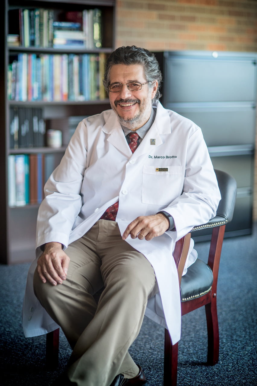
The award from the NIH’s National Institute of Dental and Craniofacial Research is the first round of funding Varanasi has received for this work since arriving at UTA in 2018. The project has been in motion since he was at his previous institution, working in collaboration with Marco Brotto, the George W. and Hazel M. Jay Professor in the College of Nursing and Health Innovation, and Pranesh Aswath, professor of materials science and engineering, both of UTA.
Brotto and Aswath are co-investigators on the NIH-funded project. Varanasi said they bring essential expertise. Aswath strengthens Varanasi’s principal understanding of materials, while Brotto, who heads the Bone-Muscle Research Center, leads the team in exploring the biological implications and experiments.

“This project represents strong collegiality and collaboration,” Varanasi said. “Not only are we bringing together some of the foremost experts at UTA, but we’ve also partnered with Simon Young at UT Health Science Center in Houston to conduct the clinical translational testing, while engaging an adviser, Harry Kim, from the Texas Scottish Rite Hospital for Children, which opens the door for additional research in applications of hip disorder.
“We’ve assembled a powerhouse team, which makes this project really exciting.”
The antioxidant material that acts as a regenerative agent when printed directly onto the bone is developed in UTA’s Shimadzu Institute Nanotechnology Research Center. Silicon oxynitride has natural antioxidant properties, as demonstrated in Varanasi’s work.
“Silicon oxynitride is derived from a material commonly used in semiconductors and microchips built for powering phones and computers,” Varanasi said. “It is intended to promote faster adhesion of bone to an implant surface when the material is used as an implant coating.”
The team believes this project could have implications for healing fractures in aging adults because of the antioxidants’ anti-aging properties, as well as potential applications for healing muscle loss—a concept that has already received federal and private inquiries for potential use in clinical applications.

“The work of this team has the potential to transform the way we approach bone health and healing,” said Aswath, who also serves as UTA’s vice provost for academic affairs. “We are combining the power of multiple disciplines to improve the quality of life for patients with craniofacial injuries.”
Varanasi added that he is most excited that his team of students, who play a critical role in his lab, are able to participate in the work.
“Our work is important, but what really matters here is that we’re engaging the next generation of scientists and setting them up with invaluable experience,” Varanasi said.